

JAKARTA - A doctor from the George Washington University Hospital managed to visualize the lungs of a patient infected with the corona virus (SARS-CoV-2) or COVID-19. This visualization is made using VR (Virtual Reality) technology developed by Surgical Theater.
Launching from CNN International, the results of the rendering of the lung organs were made by Dr. Keith Mortman from a COVID-19 positive patient. Mortman used CAT (Computer Aided Tomography) or CT scans of the patient as his database.
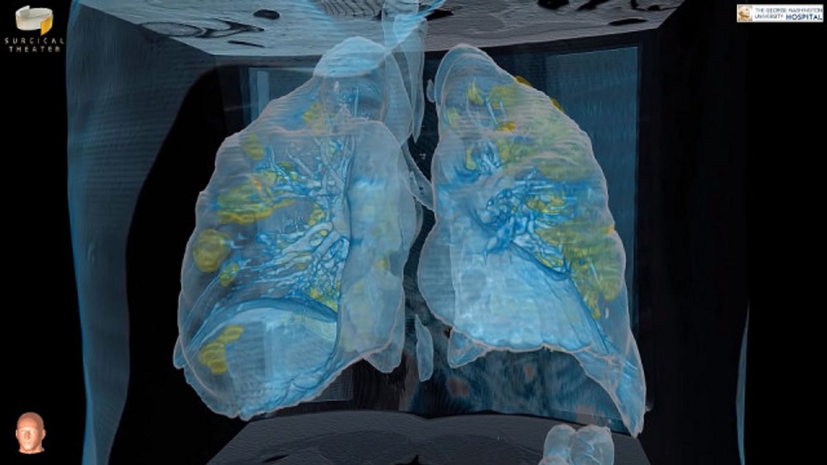
The medical record data is then processed in a VR display, and shows damage to the lung organs of the corona positive patient. Through the VR recording, the patient's condition can be monitored, wherein he needs a ventilator to help him breathe.
"It's surprising because it's not like pneumonia which might affect only a small part of the lung or not like the common cold, what you see in this video is actually a broader range of lung damage," Dr. Mortman is also chief of thoracic surgery at George Washington Hospital.
Furthermore, Mortman explained in the VR video showing the base of normal lungs in blue. However, there are many parts of these organs that are yellow, that is a sign that the corona virus has spread.
The yellow sign in the video is a viral infection and inflammation in the lungs, which is why too many of the COVID-19 patients have difficulty breathing or are short of breath. In these symptoms the patient needs to be hospitalized, wear a breathing tube, or put on a ventilator.
"The areas highlighted in yellow on the video represent parts of the lung that are infected and inflamed. From the scans, it is clear that we know that the damage is not localized to a single area, but instead covers large swaths of both lungs and shows how quickly and aggressively the infection can persist. , "continued Dr. Mortman.
He stated that once the lungs were damaged at this rate, the respiratory system in humans could take a long time to heal. Allegedly, about two to four percent of patients who test positive for COVID-19, the damage is irreversible and they will succumb to the disease.

George Washington University Hospital typically uses CT imaging technology that produces video to record human cancer and plan operations. But for the first time, the technology has now been applied to combat the COVID-19 virus.
According to Dr. Mortman, this video rendering has a strong message for the public, to stay at home, not to travel without wearing a mask and to take care of your health. Also avoid physical contact with other people.
"I really want them to be able to see this and really understand the damage that's being done to the lungs. The severity of the disease caused by this. So maybe, maybe they think twice before having a party at home or going out and hanging out with them. friends, "explained Dr. Mortman.
The English, Chinese, Japanese, Arabic, and French versions are automatically generated by the AI. So there may still be inaccuracies in translating, please always see Indonesian as our main language. (system supported by DigitalSiber.id)
Most Popular Tags
#Prabowo Subianto #donald trump #2026 World Cup #venezuela #konflik timur tengahPopular


