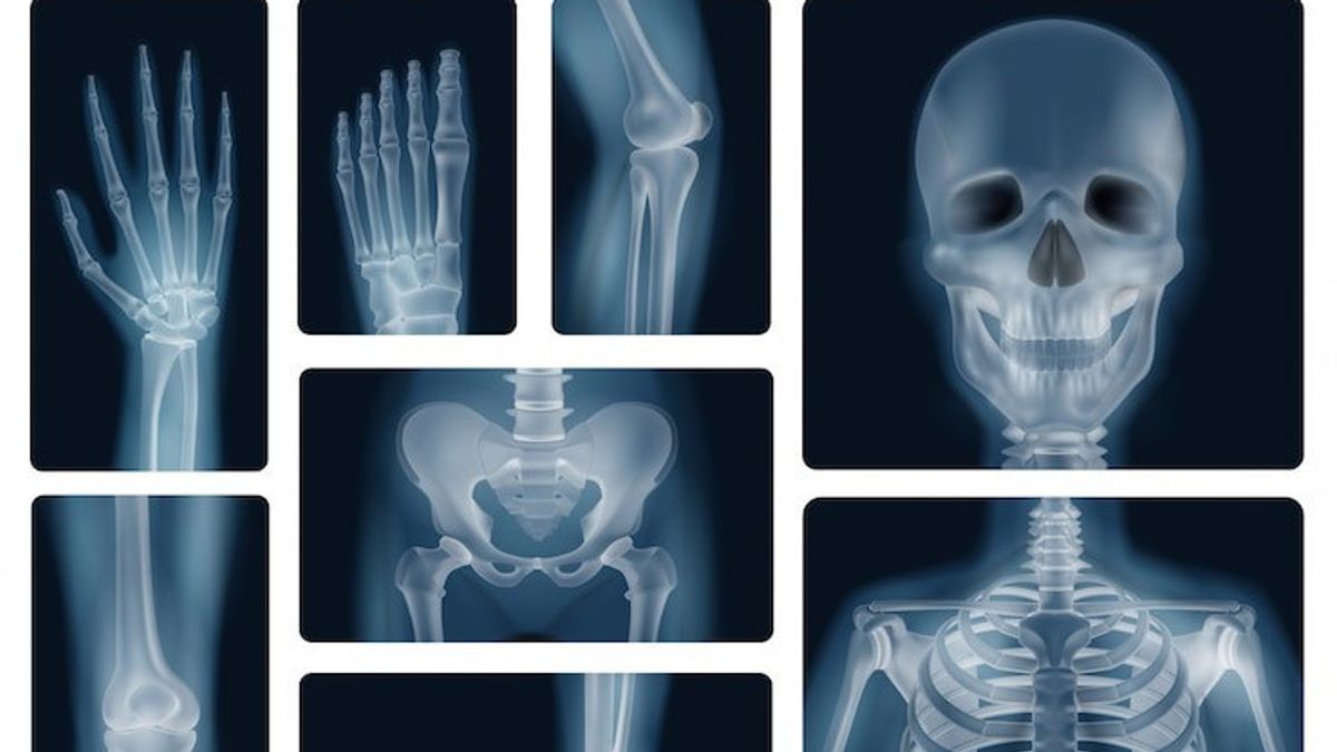
YOGYAKARTA - Are you curious about how the process of forming a bone is so hard and sturdy?
The process of bone formation, or self-osification, is the foundation of bone health. By understanding this process, we can take appropriate steps to prevent bone problems such as osteoporosis.
Reporting from the US Department of Health and Human Services page, osteogenesis and osteogenesis are often used alternately to show the process of bone formation.
The process begins when the skeleton parts form in the first few weeks after conception. At the end of the eighth week after conception, the skeleton pattern forms in the prone bone and membrane of the ikat tissue, then the ossification begins.
SEE ALSO:
The development of the bone continues into adulthood. Even after the adult height is achieved, bone development continues to repair fractures and remodelize according to changing lifestyle needs.
Also read the article that discusses 5 Safe Sports Tips for Osteoporosis Sufferers
Osteoblas, osteocyt, and osteoclas are three types of cells involved in bone development, growth, and remodelation. Osteoblas is a cell that forms bones, osteocyt is an adult bone cell, and osteoklas destroys and reabsorbs the bone.
Intramembranosa Osification involves replacing the membrane of a sheet-shaped braid tissue with a bone tissue. The bone formed in this way is called the intramembranosa bone.
For example, there are several flat bones on the skull and several other irregular bones. Future bones were first formed as membranes of ikat tissue. Osteoblas migrated to the membrane and deposited a bone matrix around it. Then when the osteoblast is surrounded by a matrix it will be called an osteosit.
Endokondral's Osification involves replacing hialin-prone bones with bone tissue. Most of the skeleton bones are formed in this way and are called endokondral bones.
In this process, future bones are first formed as models of hialin-prone bones. Then in the third month after fertilization, the pericondrium that surrounds this cartilage model becomes affiliated with blood vessels and osteoblas, then turns into a periodium.
Osteoblas formed a compact bone layer around the diaphysis. At the same time, the cartilage in the middle of the diaphysis begins to decompose. Osteoblas penetrated the broken bones and replaced them with sponge bones, forming a primary ossification center. Osification continued from this center towards the ends of the bones.
Then after the sponge bone is formed in diaphysis, osteoclas destroys the newly formed bone to open a meduler cavity. The susceptible bone in epiphysis continues to grow so that the growing bone increases.
Then, usually after humans are born, secondary oscism centers form in epiphysics. The oscive process in epiphysis is similar to in dialysis, but sponge bones are preserved and not destroyed to form a meduler cavity.
When secondary ossification is complete, the hialin-prone bone is completely replaced by bone, except in two areas: one area remains on the epiphysics surface as an articularly prone bone, and the other region remains between epiphysics and diaphosis as an epiphysic plate or a growth area.
Tulang kemudian akan tumbuh melongjang di lempeng epifisis melalui ossifikasi endokondral hingga masa 20-an, diatur Hormor pertumbuhan dan seksex.
After stopping to lengthen, the bone can still gain thickness (abelitional growth) due to osteoblas and osteoklas working together, responding to stress due to muscle activity or body burdens.
In addition to the process of bone formation, follow other interesting articles too. Want to know other interesting information? Don't miss it, keep an eye on the updated news from VOI and follow all of its social media accounts!
The English, Chinese, Japanese, Arabic, and French versions are automatically generated by the AI. So there may still be inaccuracies in translating, please always see Indonesian as our main language. (system supported by DigitalSiber.id)


